health journal

I   Home    I    firstaid   I    Nurses recruitment   I    Medical Equipment   I    Hospital Supplies   I    Dialysis Machine   I    Ultra Sound   I    First Aid   I    Cancer   I    Contact   I    Career   I


Wednesday, August 23, 2006

Obesity


Obesity

By; Sandi Effendi
urology staff nurse mubarak al kabeer hospital, kuwait

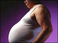
Obesity is an overabundance of body fat resulting in body weight of 20% or more than the average weight for the person’s age, height, sex, and body frame. Increasingly, obesity is being diagnosed using the Body Mass Index (to account for body build) and/or Body Surface Area and Basal Metabolic Rates (to account for metabolic activity for the person). A BMI greater than 30 is considered obese. About 18% of American are obese (up from 12% in 1991) and 63& of men and 55% of women are over weight.

Pathophysiology and etiology
1. Increasing evidence reveals that heredity plays a part in the development of obesity. Identical twins raised apart are more likely to have similar amounts of body fat than fraternal twin raised separately.
2. Environment plays a role.
a. Some evidence shows that children reared by obese parents have an increased tendency toward obesity.
b. In addition, social class may be associated with more weight-conscious behavior.
3. A variety of psychological factors may contribute to weight gain, including depression and anxiety.
4. Physiologic factors
a. Endocrine abnormalities (rare causes of obesity)-Cushing’s syndrome, hypothyroidism, hypogonadism, or hypothalamis lesions.
b. Age-advancing age may be associated with obesity often because of changes in activity level or in women, because of hormonal changes; early childhood and the start of puberty may also be associated with obesity.
(i) Overeating after puberty may increase the total number of fat cells.
(ii) Despite dieting, these extra ft cells can never be eliminated; they only decrease in size.

Clinical Manifestation
1. Body weight greater than 20% of acceptable weight for eight or BMI > 30.
2. Increased weight is correlated with increased incidence of:
a. Cardiovascular disease
b. Diabetes mellitus

Diagnostic evaluation
Conservative Measures
1. Diet therapy-has been controversial, but a well-balanced diet containing all the major food groups is still advised.
a. One thousand calories per day must be eliminated from a diet to lose 1 kg (2.2 lb) of body weight per week.
b. A 1,200-calorie diet for women and a 1,500-calorie diet for men with variations depending on patient size and activity level are basic to diet management. Fats should compose no more than 30 % of all calories, proteins approximately 15 to 20% and carbohydrates should constitute the remaining portion
c. A balance of food groups is essential to maintain vitamin and nutrient balance. Nutrient supplements may be necessary (iron, B6,Zinc, and folate)
d. Food preparation, should include seasoning with herbs, onion, garlic, and pepper, and foods should be baked, broiled, steamed, or sauteed using minimal polyunsaturated oil.
e. Food attractively arranged on smaller plates, using whole rather than processed foods and eaten slowly, will assist the overall process
f. Eliminating entire food groups from the diet, such as carbohydrates (in many popular protein and fat-based diets), will eventually result in craving of those foods eliminated, disruption of normal metabolic processes, and quick weight gain when the food is added to the diet.
2. Exercise—a daily exercise program may include walking or other aerobic activities for approximately 180 minutes per week, or 1 hour at least three times a week, however daily exercise is optimal.
3. Behavior modification is a cornerstone of any successful diet.
a. Identify and eliminate situations or cues leading to overeating or high-calorie foods with use of a food diary.
b. Provide positive reinforcement of proper dietary habits.
c. Should a lapse in diet habits occur, focus on a prompt and positive return to appropriate dietary habits
d. Stress reduction techniques, such as visual imagery or progressive relaxation; peer support may be helpful

Pharmacotherapy
1. Anorexia medications, such as amphetamines and norepinehrine-releasing agents or reuptake inhibitors, reduce appetite and stimulate weight loss initially.
2. However, tolerance develops within 2 to 4 weeks and weight is rapidly regained when the drugs are discontinued
3. Numerous long-term studies have failed to show long term success with these agents
4. Phentermine (Ionamin, Fastin) is one of the most widely prescribed agents; however, it causes stimulating effects and should not be used in uncontrolled hypertension, advanced heart disease, history of drug abuse, and with MAO inhibitors.
5. Sibutramine (Meridia) is a mixed neurotransmitter reuptake inhibitor that acts on the central nervous system to reduce appetite.
a. The long-term risks of the medication are not known, but it must be used cautiously with hypertension, coronary artery disease, heart failure, arrhythmia, renal and hepatic impairment, narrow angle glaucoma, and seizure disorders.
b. Multiple drug interactions include monamine oxidase inhibitors, other serotonic drugs (selective serotonin reuptake inhibitors (SSRIs) antidepressants, sumatriptahn and other migraine agents), lithium, dextromethorphan, and possible erythromycin and ketoconazole.
c. Side effects include dry mouth, constipation, dizziness, nervousness, insomnia. Has not been shown to be addictive.
6. Recently, scientists have found success in weight loss with the use of orlistat (xenical), agastrointestinal lipase inhibitor, which blocks the breakdown of fat in the GI system. About 30% of dietary fat is eliminated.
a. Side effects include oily or fatty stools, flatulence, and GI distress.
b. Long term safety of the drug has not been determined, but addition of fat-soluble vitamin supplement (vitamin A,D,E,K and beta carotene) taken prevent a theoretical vitamin deficiency.
c. Should not be used in cases of cholestasis or malabsorption.
d. These medications are only adjunct to diet and exercise therapy.

Surgical Interventions
Numerous surgical procedures have been used. However, gastroplasty is the current procedure of choice. These therapies are generally reserved for morbidity obese patients who cannot lose weight through the above therapies.
1. Gastroplasty-most common procedure is vertical banding involving creation of a 30 ml pouch along the lesser gastric curvature with a small outlet created with the use of a ring of plastic at the distal end to prevent dilation
2. Gastric bypass-a Roux-en-Y gastroenterostomy is constructed by first creating a 50 –ml pouch in the proximal stomach by stapling horizontally and completely separating the smaller proximal stomach pouch from the larger distal stomach pouch. To this proximal pouch, the distal jejunum is attached, thus bypassing the distal stomach pouch. The transected proximal portion of the jejunum is anastomosed to the distal jejunum.

Complications
1. Obesity is a risk factor for diabetes, gallbladder disease, osteoarthritis of weight-gearing joints, high blood pressure, and coronary artery disease.
2. Vitamin and mineral deficiencies because of surgical intervention and/or severely restricted diet
a. A moderate, well-balanced weight reduction diet will generally not cause deficiencies, although a multiple vitamin/ mineral supplement may be used
b. A low-calorie diet (fewer than 800 to 1,000 calories/day) will require careful monitoring and vitamin/ mineral supplements.

Nursing Assessment
1. Obtaining a complete nutritional assessment (may be in collaboration with a nutritionist)
2. Assess behavioral/ emotional components of eating, coping mechanism, and past successes/ failures with dieting.

Nursing diagnosis
 Altered Nutrition: More Than body Requirements related to high-calorie, high-fat diet, and limited exercise
 Fluid volume Deficit related to gastroplasty or gastric bypass surgery
 Self esteem Disturbance related to weight

Nursing Interventions
Modifying Nutritional Intake
1. Assist patient in assessing current dietary habits and identifying poor dietary habits
2. Assist patient in developing appropriate diet plan based on likes and dislikes, activity level, and lifestyle
3. Suggest behavior modification strategies, such as shortening lunch break, preventing access to quick snacks, eating only at mealtimes at the table
4. Provide emotional support to patient during weight reduction efforts positive reinforcement and creative problem solving
5. Provide patient with alternative coping mechanisms, including stress reduction techniques, such as progressive relaxation and guided imagery
6. Assess patient’s ability to tolerate exercise through measurement of vital signs before, during, and after exercise and ask about symptoms of shortness of breath and chest pain

Outcome Based Evaluation
 Five pound weight loss during first month
 No abdominal distention, nausea, vomiting, or wound infection
 verbalizes feeling good about self secondary to change in diet exercise habits

Source reference

The Lippincott seventh edition

Eczema

Eczema
(infantile and childhood eczema)

by; Sandi Effendi
urology staff nurse in mubarak al kabeer hospital kuwait

Atopic dermatitis the most common cause of eczema in childhood, is a characteristic inflammatory response of the skin. The major problem features include pruritus, a typical
Morphology and distribution, a chronic or chronically relapsing nature, and personal or family history of atopy (asthma, hay fever, and atopic dermatitis). There is tendency toward dry skin and lower threshold of itching.
Atopic dermatitis affects 10 % to 15% of the childhood population. It usually starts after 2 months of age. By age 5, 90% of the patients who will develop atopic dermatitis have already manifested the disease. It may stop after an indefinite period of time, or it may progress from infancy to adulthood with little or no relief. It is rare of adults to develop atopic dermatitis without a history of eczema in childhood.

Pathophysiology and Etiology
1. Atopic dermatitis involves immunologic abnormalities, such as elevated immunoglobulin E levels and increased rates of sensitization to common contact allergens and to intradermal skin tests. Although the exact cause is unknown, there is a constitutional predispotition to develop pruritus. In general, the skin of patients with atopic dermatitis is different from that of healthy patients in the following respects:
a. Increased tendency toward dryness
b. Lowered threshold for pruritis from minor irritants, such as soap, perspiration, cold weather, and heat
c. Tendency toward lichenification (leathery thickening of skin ) and production of a rash when the skin is rubbed or scratched
2. The etiology is unknown but has familial tendencies. Almost 75 % of patients with this form of eczema will develop hay fever or asthma themselves.

Clinical manifestations
Age and Distribution of the lesions
Atopic dermatitis is divided into three phases based on the age of the patient and the distribution of the lesions. There are referred to as the infant, childhood, and adult phases.
1. Infant (2 months to 2 years):
a. The onset is between 2 and 6 months of age. Half of affected infants have spontaneous resolution by age 2 or 3
b. Characterized by intense itching, erythema, papules, vesicles, oozing, and crusting
c. The rash usually begins on the cheeks, forehead, or scalp and then extends to the trunk or extremities in scattered, often symmetric patches. The perioral, perinasal, and diaper areas are usually spared
2. Childhood (4 to 10 years):
a. Affected persons in this age group are less likely to have exudates and crusted lesions. Eruption are characteristically more dry and popular and often occur as circumscribed scaly patches. There is a greater tendency toward chronicity and lichenification
b. The typical areas of involvement are the face, including the perioral and perinasal areas, neck, antecubital and popliteal fossae, wrists, and ankles.
3. Adult (puberty to old age):
a. Predominant areas of involvement include the flexor folds, face, neck, upper arms, back, dorsa of the hands and feet, fingers, and toes.
b. The eruption appears as thick, dry lesions, confluent papules, and large lichenified plaques. Weeping, crusting, and exudation can occur, but they are usually the result of superimposed external irritation or infection.

Clinical Appearance
Atopic dermatitis is also divided into three stages based on the clinical appearance of the lesions. The acute, subacute, and chronic stages can occur in infants, children, and adults,
1. Acute moderate to intense eryhema, vesicles, a wet surface, and severe itching.
2. Subacute:
a. Erythema and scaling are present in various patterns with indistinct borders. The redness may be faint or intense. The surface is dry. There are varying degrees of pruritus.
b. The subacute stage may be an initial stage or may follow an acute inflammation or exacerbation of a chronic stage. Irritation, allergy, or infection can convert a subacute process into an acute one.
3. Chronic the inflamed area thickens and the surface skin markings become more prominent. Thick plaques with deep parallel skin markings are called lichenified. Lichenification is the hallmark of chronic eczema. The surface of the skin is dry and the border of the lesion well defined. There is moderate to intense itching.

Diagnostic Evaluation
Atopic dermatitis is usually a clinical diagnosis based on the evaluation of the aggregate of signs, symptoms, stigmata, course, and associated familial findings. The major features include pruritus, a characteristic morphology and typical distribution for the age of the patient, a chronic or chronically relapsing nature, and a personal or family history of atopical disease. When the diagnosis is in doubt, a skin biopsy may be performed.

Treatment
Acute
1. Open wet dressing for 1 to 3 days
2. Avoidance of any known allergen
3. Topical corticosteroids
a. Topical corticosteroids are ranked into seven groups according to potency. Group 1 contains the most potent topical steroids and group 7 the least potent ones. The concentration listed on the medication does not correlate with its potency or safety, but is merely a statement of its specific chemical formulation.
b. Very potent (Group 1) topical corticosteroids are avoided in children younger than 12 years of age because of greater skin absorption.
4. Oral medication to relieve itching- hydroxyzine hydrochloride (ataraz), diphenhydramine hydrochloride (Benadryl), or protmethazine hydrochloride (Phenergan). Diphenhydramine and promethazine cause more sedation than hydroxyzine hydrochloride. Mild sedation may be desirable.
5. Management of secondary infection, if present, with oral antibiotics
6. Initiation of a hypoallergenic diet to eliminate any responsible food for infants and young children with severe, recalcitrant atopic dermatitis
Subacute and Chronic
1. Prevention of dry skin:
a. Diminish the frequency and duration of bathing
b. Use mild soap and hydrophilic lotion
c. Lubricate the skin with emollients
d. Add tar preparations to the bath water
e. Maintain environmental humidity above 40% in winter months
2. Same measures as acute stage with exception of wet dressings

Nursing Assessment
1. Take a nursing history focusing on clinical manifestations:
a. Onset and duration of rash
b. Location, course, and distribution of lesions
c. Change in morphology of lesions
d. Local and systemic symptoms
e. Exposure to possible allergens
f. Previous episodes of rashes
g. Personal history of allergies, asthma, or hay fever
h. Family history of eczema, allergies, or hay fever
i. Medications, treatments tried, and their effect
2. Perform a physical assessment.
a. Examine the entire skin in an orderly fashion with specific attention to the type of lesion (ie, macule, papule, vesicle, etc), its appearance (shape, border, color, texture, and surface), and its distribution (areas of the body involved)
b. Note any associated symptoms, such as scratching, fever, or drainage.
3. Document findings:
a. Describe skin finding using dermatologic terminology
b. Draw pictures to facilitate communication.

Nursing Diagnoses
 Impaired skin Integrity or high risk for impairment, related to skin pathology and scratching
 Sensory/ Perceptual Alterations (Tactile), related to skin pathology
 Risk for infection related to increased bacterial colonization of skin and possible break in defensive barrier

Nursing Interventions
The nurse may perform the following interventions or teach the patient or family to do the following:
Improving skin integrity
1. Reduce inflammation during the acute stage with the topical application of open wet dressings.
a. Use a soft, lightweight cloth, such as a handkerchief, a thin diaper, or strips of bed sheeting. Do not use gauze (adheres to skin), washcloths, or towels (too heavy)
b. Open wet dressings should be clean. In certain situations, they should be sterile to prevent contamination.
c. Solutions should be lukewarm or at body temperature to soothe the skin and prevent chilling.
d. Compresses should be moderately wet, not dripping, and removed after 20 minutes, unless otherwise directed. They should be reapplied three to four times a day.
e. After the compress, a topical corticosteroid may be applied to further reduce itching and inflammation.
f. Observe the skin for changes in response to therapy
2. Prevent dry skin during the subacute and chronic stages.
a. Decrease the frequency and duration of bathing, long, hot tub are not be avoided.
b. Avoid hot water and harsh soaps. Patients should bathe in lukewarm water using mild soap (dove, Neutrogena); avoid bubble baths ;rinse well and pat skin dry with towel.
c. If bath water stings, add 1 cup of table salt.
d. Apply unscented emollients (eg, Eucerin, Keri, Lubriderm) within 3 minutes of bathing, when the skin is slightly moist. Creams and ointment are more effective than lotions because they are better at preventing evaporation of water from the skin. Bathing will dry and damage skin unless an emollient is applied immediately after existing bath.
e. Some patients may benefit from soaking in a tar bath for 15 to 20 minutes daily, preferably in the evening. Add to bath skin and clothing and may cause sunlight sensitivity.
f. For patient with extremely dry skin, cleanse with a hydrophilic lotion (eg. Cetaphil). Apply without water until light foam occurs. Remove by wiping with soft cotton cloth or cleansing tissue.
g. Keep environmental humidity above 40 % in winter months. Use a humidifier.
h. Observe the skin for changes in response to therapy.

Controlling Pruritus
1. Apply topical corticosteroids.
a. Apply a thin layer of topical corticosteroids to the affected skin two to four times a day as directed. Use only for the duration prescribed.
b. Observe for possible side effects from long-term use of topical corticosteroids (ie, striae, cutaneous atrophy, telangiectasia, acne, and growth retardation).
c. Note any scratching and intervene as necessary
2. Administer oral antipruritic medications
a. Give medications exactly as prescribed
b. Note the degree of sedation and presence of scratching.
3. Teach the care taker or family of infants and small children a hypoallergenic diet when indicated.
a. Write any known allergens on care plan and chart. Inform the dietitian of the child’s food allergies
b. Avoid substances that have a high potential for sensitization, such as cow’s milk, eggs, tomatoes, citrus fruits, chocolate, wheat products, spiced food, fish, nuts, and peanut butter.
c. A minimal diet is prescribed. The trial diet may be composed of milk substitute, rice cereal, two fruits, two vegetables, beef, a multivitamin, and no eggs.
d. A new food is added to the diet every 3 to 5 days, during which time the response to the food is observed.
e. An allergic response occurring during this 3 to 5 day period indicates sensitivity to that food. The food is then eliminated from the diet. If no response is apparent, the food is added to the child’s diet.
f. Another food substance is then added, and child is observed for the following 3 to 5 days period. This method is followed until the food allergen is determined.

Preventing infection
1. Assess and/or treat secondary infection:
a. Observe the skin for sign of bacterial infection (discharge, oozing, crusts). Report positive findings
b. Administer antibiotic as prescribed.
c. Loosen exudates and crusts with water or wet dressings, unless otherwise specified.
d. Note changes in the skin in response to therapy

Selected references
Boiko,S (200), Making rash decisions in the diaper area. Pediatric annals
Habif, T.P. (1996), Clinical dermatology: A color guide to diagnosis and therapy
Hall, J.C. (2000) Sauer’s manual of skin diseases. Philadelphia: Lippincott Williams and Wilkins.

Tuesday, August 22, 2006

Hydronephrosis

By:
Sandi Effendi
staff nurse in urology ward, mubarak al kabeer hospital kuwait

Hydronephrosis is dilation of the renal pelvis and calyces of one or both kidneys due to an obstruction.

Pathophysiology
Obstruction to the normal flow of urine causes the urine to back up, resulting in increased pressure in the kidney. If the obstruction is in the urethra or the bladder, the back pressure affects both kidneys, but if the obstruction is in one of the ureters because of a stone or kink, only one kidney is damaged.
Partial or intermittent obstruction may be caused by a renal stone that has formed in the renal pelvis but has moved into the ureter and blocked it. The obstruction may be due to a tumor pressing on the ureter or to bands of scar tissue resulting from an abscess or inflammation near the ureter as it leaves the renal pelvis or to an unusual position of the kidney, favoring a ureter obstruction at the elderly men, the most common cause is ureteral obstruction at the bladder outlet by an enlarged prostate gland. Hydronephrosis can also occur in pregnancy because of the enlarged uterus.
Whatever the cause, as the urine accumulates in the renal pelvis, it distends the pelvis and its calyces. In time, atrophy of the kidney results. As one kidney undergoes gradual destruction, the other kidney gradually enlarges (compensatory hypertrophy). Ultimately, renal function is impaired.

Clinical manifestations
The patient may not have symptoms if the onset is gradual. Acute obstruction may produce aching in the flank and back. If infection is present, dysuria, chills, fever, tenderness, and pyuria may occur. Hematuria and pyuria may be present. If both kidneys are affected, signs and symptoms of chronic renal failure may develop.

Medical management
The goal of management are to identify and correct the cause of the obstruction, to treat infection, and to restore and conserve renal function. To relieve the obstruction, the urine may have to be diverted by nephrostomy or another type of diversion. The infection is treated with antibiotic agents because residual urine in the calyces leads to infection and pyelonephritis. The patient is prepared for surgical removal of obstructive leasions (calculus, tumor, obstruction of the ureter). If one kidney is severely damaged and its function is destroyed, nephrectomy (removal of the kidney) mat be performed.

Friday, August 18, 2006

MIGRAINE

Migraine is a neurologic disease, of which the most common symptom is an intense and disabling headache. Migraine is the most common type of vascular headache. Migraine headaches are usually characterized by severe pain on one or both sides of the head, an upset stomach, and at times disturbed vision. The word "Migraine" comes from the Greek construction "hemikranion" (ημικρανίον, pain affecting one side of the head)
Signs & Symptoms

Migraine with aura is a neurological disease characterized by flare-ups generally referred to as "Migraine attacks." "Aura" refers to the non-headache features of Migraine that often happen before, or in the place of, the actual headache.

It is possible to have a Migraine attack marked by other symptoms and no headache at all, which is called acephalgic Migraine. The aura symptoms associated with these Migraines are very similar to the symptoms of Chronic Fatigue Syndrome, and thus it is possible that the two may be related, or even the same.

Many Migraine sufferers have headache without aura. Such headaches are commonly misdiagnosed as sinusitis or chronic sinus infections. The error can be revealed by a CT scan of the sinuses, which will show inflammation in the sinuses if a sinus infection is present. Migraine had been thought to be caused by vasodilation in the head and neck; however, newer research suggests that vascular dilation associated with Migraine is a symptom of Migraine, not the cause of Migraine symptoms.

Blood vessel diameter is under neurochemical control; in other words, blood vessels dilate during a Migraine episode because the nervous system tells them to. The cause of the pain itself is from activation of the trigeminal nerve. This theory is still being examined though. The trigger of the Migraine may be overactivity of nerve cells in certain areas of the brain (for example, the raphe nucleus). Often a Migraine episode is associated with strong emotional expression or psychic tension, but those may be Migraine symptoms rather than Migraine triggers.

The pain from a Migraine is typically one-sided, though it may encompass the whole head, or move from side-to-side as the Migraine progresses. Additionally, the pain from a Migraine is usually described as throbbing and moderate to severe in intensity. Migraines are frequently accompanied by nausea/vomiting and either photophobia (excessive sensitivity to light) or phonophobia (excessive sensitivity to sound), causing the sufferer to seek a dark, quiet room for recovery. If the predominant feature is vomiting or nausea cyclic vomiting syndrome should be considered as a possible cause.

In Migraine with aura, formerly called classical Migraine, the headache phase is preceded or accompanied by a group of specific symptoms called aura, most commonly experienced as a visual disturbance prior to the attack. Aura usually lasts less than 60 minutes, and in those who suffer Migraine with aura there is generally little time between the onset of aura and the onset of the attack. Migraine without aura, formerly called common Migraine, in contrast to Migraine with aura, lacks any manifestations associated with headache. Some experience aura without Migraine, a condition formerly called amigrainous Migraine or optical Migraine, now usually called acephalgic Migraine. Although sometimes comparable in severity, the symptoms of Migraine differ from those of cluster headache.

Visual aura can include castellated scotoma or fortification spectra, multicolored zig-zag patterns which can cover a large part of the visual field of one eye (sometimes both). Other types of visual aura involve distortions in perception of color, such as color bleeding or the appearance of halos, or as a white spot in the visual field, similar to when a camera flash temporarily "blinds" your vision.

While the most common type of aura is visual, it can manifest as any specific neurological symptom complex. Some experience tingling sensations called paresthesias or disturbances of other regions of the brain (such as language ability or smell) instead of a visual aura, either as an occasional alternate or as their normal aura. Some experience unusual odors that are not actually present, fatigue, nausea, balance problems, and vertigo.

Aura need not be related to the five senses: many Migraineurs experience a prodrome, a vague feeling that things are just not right. While the types and severity of aura can be extremely diverse, a given sufferer will generally experience similar manifestations of aura with each Migraine attack. Many people experience difficulty in speaking and/or forming cohesive syntax.

Migraine can accompany, in many cases, another type of headache called tension headache. Studies have demonstrated that, in patients who get both Migraines and tension headaches, their tension headaches will respond to their usual Migraine treatment. This is in contrast to patients who get only tension headaches.

Because chronic tension headaches are sometimes caused by the same triggers as Migraine and can often be remedied using the same treatments used for Migraine, and because Migraine itself presents with different symptoms for different people, there are some who believe that chronic tension headaches are just another symptom of Migraine.

Migraines can be associated with seizures. Stroke symptoms are seen in some patients and are known as complicated Migraine; these symptoms should not be permanent.

Migraine often runs in families and starts in adolescence, although evidence indicates that it starts also in childhood (including infants) or even in utero. In children, Migraine has some distinct features: headache is more often bilateral or difficult to localize, the patient is unable to describe the symptoms with significant accuracy; in infants, Migraine attacks may be manifested by periods of somnolence or irritability.

Because their symptoms vary, an intense headache may be misdiagnosed as a Migraine by a layperson. Indeed, many other headaches, some of them caused by very serious diseases (like a brain tumour, hydrocephalus, brain vascular disorders) may have a great resemblance to the clinical picture of Migraine and can lead to misdiagnosis. Where possible, see a doctor to determine if the headaches are a symptom of something else.

Hemiplegic Migraine may have periods of one-sided paralysis. This is one of the few types with a known etimology, a defect in atomic-size pores in the cell membranes of nerves that admit calcium into the cell. It may be associated with loss of conscioiusness and is often misdiagnosed as epilepsy. It can often be effectively prevented with medicines called calcium channel blockers.

A less frequently seen type of Migraine is the Basilar Type Migraine, which, until recently had also been referred to as a Basilary Artery Migraine. These Migraines have many of the same symptoms as Migraine with aura or Migraine without aura, but affect a different part of the brain, and are oftentimes accompanied by difficulty walking, speaking, or use of other motor skills. Other symptoms of Basilar Type Migraine may include depersonalization, auditory and visual hallucinations, and a distorted time sense.


Pathophysiology

Research scientists are unclear about the precise cause of Migraine headaches. There seems to be general agreement, however, that a key element is blood flow changes in the brain. People who get Migraine headaches appear to have blood vessels that overreact to various triggers.

Scientists have devised one theory of Migraine which explains these blood flow changes and also certain biochemical changes that may be involved in the headache process. According to this theory, the nervous system responds to a trigger such as stress by causing a spasm of the nerve-rich arteries at the base of the brain. The spasm closes down or constricts several arteries supplying blood to the brain, including the scalp artery and the carotid or neck arteries.

As these arteries constrict, the flow of blood to the brain is reduced. At the same time, blood-clotting particles called platelets clump together-a process which is believed to release a chemical called serotonin. Serotonin acts as a powerful constrictor of arteries, further reducing the blood supply to the brain.

Reduced blood flow decreases the brain's supply of oxygen. Symptoms (neurological symptoms) signaling a headache, such as distorted vision or speech, may then result, similar to symptoms of stroke.

Reacting to the reduced oxygen supply, certain arteries within the brain open wider to meet the brain's energy needs. This widening or dilation spreads, finally affecting the neck and scalp arteries. The dilation of these arteries triggers the release of pain-producing substances called prostaglandins from various tissues and blood cells. Chemicals which cause inflammation and swelling, and substances which increase sensitivity to pain, are also released. The circulation of these chemicals and the dilation of the scalp arteries stimulate the pain-sensitive nociceptors. The result, according to this theory: a throbbing pain in the head.

More recent imaging techniques seem to show that Migraine is primarily a disorder of the brain (neurological), not of the blood vessels (vascular). A spreading depolarization (electrical changes) may begin 24 hours before the attack, with onset of the headache occurring at about the time of maximum brain coverage. The effects of Migraine may persist for some days after the main headache has ended. Many sufferers report a sore feeling in the area where the Migraine was, and some report impaired thinking for a few days after the headache has passed.

In 2005, research was published indicating that in some people with a patent foramen ovale (PFO), a hole between the upper chambers of the heart, Migraine might result and that the Migraine ended if the hole were blocked. That procedure is hazardous enough that it may not be wise to treat solely to prevent Migraine, but if there is evidence of small strokes on magnetic resonance imaging, the procedure could be worthwhile. Early speculation as to the cause of the relationship has centered on the idea that the lungs detoxify blood as it passes through. The PFO allows blood to go directly from the right side of the heart to the left without passing through the lungs.


Migraine triggers

Migraine is irregularly episodic, so there needs to be some explanation for why a particular Migraine episode occurs at a particular time and not at another time.

The trigger theory supposes that exposure to various environmental factors precipitates, or triggers, individual Migraine episodes. Many people report that one or more dietary, physical, hormonal, emotional, or environmental factors precipitate their Migraines. The most-often reported triggers include stress, alcohol, foods, too much or too little sleep, and weather. Sometimes the Migraine occurs with no apparent "cause".

Migraine patients have long been advised to try to identify personal headache triggers by looking for associations between their headaches and various suspected trigger factors. Patients are urged to keep a “headache diary” in which to note what they eat and when they get a headache, to look for correlations, and to try to avoid headache by avoiding factors they identify as triggers. Typically this advice is accompanied by a list of trigger factors.

Authors who in 2005 reviewed the medical literature found that the available information about dietary trigger factors relies mostly on the subjective assessments of patients. Some suspected dietary trigger factors appear to genuinely promote or precipitate Migraine episodes, but many other suspected dietary triggers have never been demonstrated to trigger Migraines. The review authors found that alcohol, caffeine withdrawal, and missing meals are the most important dietary Migraine precipitants. The authors say dehydration deserves more attention, and that some patients are sensitive to red wine. The authors found little or no demonstrated evidence that notorious suspected triggers chocolate, cheese, or that histamine, tyramine, nitrates, or nitrites normally present in foods trigger headaches. The artificial sweetener aspartame (NutraSweet) has not been shown to trigger headache, but in a large and definitive study monosodium glutamate (MSG) in large doses (2.5 grams) was associated with adverse symptoms including headache more often than was placebo. The review authors also note that general dietary restriction has not been demonstrated to be an effective Migraine therapy.


Treatment

Conventional treatment focuses on three areas: trigger avoidance, symptomatic control, and preventive drugs. Patients who experience Migraines often find that the recommended treatments are not 100% effective at preventing Migraines.


Trigger avoidance

Patients can attempt to identify and avoid factors that promote or precipitate Migraine episodes. Moderation in alcohol and caffeine intake, consistency in sleep habits, and regular meals may be helpful. Beyond an often pronounced placebo effect, general dietary restriction has not been demonstrated to be an effective approach to treating Migraine.


Symptomatic control to abort attacks

Migraine sufferers usually develop their own coping mechanisms for intractable pain. A cold or hot shower directed at the head, a wet washcloth, less often a warm bath, or resting in a dark and silent room may be as helpful as medication for many patients, but both should be used when needed.

For patients who have been diagnosed with recurring Migraines, doctors recommend taking painkillers to treat the attack as soon as possible. Many patients avoid taking their medications when an attack is beginning, hoping that "it will go away". However in many cases once an attack is underway, it can become intensely painful, last for a long time (sometimes even for several days), and become somewhat resistant to medical treatment. In contrast, treating the attack at the onset can often abort it before it becomes serious, and can reduce the frequency of subsequent attacks in the near-term.

The first line of treatment is over-the-counter abortive medication. Doctors start patients off with simple analgesics, such as paracetamol (acetaminophen), aspirin and caffeine. They may provide some relief, although they are not effective for most sufferers. Some patients find relief from taking Benadryl or other anti-nausea agents.

Narcotic pain killers (for example, codeine, morphine or other opiates) provide variable relief, but their side effects, the possibility of causing rebound headaches or analgesic overuse headache, and the risk of addiction contraindicates their general use.

If over-the-counter medications do not work, the next step for many doctors is to prescribe fioricet or fiorinal, which is a combination of butalbital (a barbiturate), acetaminophen (in fioricet) or acetylsalicylic acid (in fiorinal), and caffeine. While the risk of addiction is low, butalbital can be habit-forming if used daily, and it can also lead to rebound headaches.

Anti-emetics by suppository or injection may be needed in cases where vomiting dominates the symptoms. The earlier these drugs are taken in the attack, the better their effect.

Until the introduction of sumatriptan (Imitrex®/Imigran®) around 1985, ergot derivatives (see ergoline) were the primary oral drugs available to abort a Migraine once it is underway. However, ergotamine tablets (usually with caffeine), though sometimes effective, have fallen out of favour. Absorption is erratic unless taken by suppository or injection. Dihydroergotamine (DHE), which must be injected or inhaled, can also be effective. These drugs can be used either as a preventive or abortive therapy.

Sumatriptan and related selective serotonin receptor agonists are now the therapy of choice for severe Migraine attacks that cannot be controlled by other means. They are highly effective, reducing the symptoms or aborting the attack within 30 to 90 minutes in 70-80% of patients. Some patients have a recurrent Migraine later in the day, and only one such recurrence in a day can be treated with a second dose of a triptan. They have few side effects if used in correct dosage and frequency. Some members of this family of drugs are:
Sumatriptan (Imitrex®, Imigran®)
Zolmitriptan (Zomig®)
Naratriptan (Amerge®, Naramig®)
Rizatriptan (Maxalt®)
Eletriptan (Relpax®)
Frovatriptan (Frova®)
Almotriptan (Almogran®)

Evidence is accumulating that these drugs are effective because they act on serotonin receptors in nerve endings as well as the blood vessels. This leads to a decrease in the release of several peptides, including CGRP and Substance P.

These drugs are available only by prescription (US and UK). Many Migraine sufferers do not use them only because they have not sought treatment from a physician.

Regarding comparative effectiveness of these drugs used to abort Migraine atacks, a 2004 placebo-controlled trial (Cephalalgia. 2004 Nov;24(11):947-54) reveals that acetylsalicylic acid, sumatriptan and ibuprofen are equally effective.

Triptan therapy has been shown to result in a reduction in lost productivity. Sumatriptan has been shown to result in an average of 0.5 fewer missed workdays during the first three months of therapy and 0.7 fewer missed workdays within the first six months, as well as a reduction in the number of days spent working while symptomatic. The average reduction in lost productivity has been estimated at $1,249, at a cost of $25 per day of disability avoided. The annual net savings in reduced health care costs and lost productivity, over the increased cost of triptan therapy, has been estimated at between $114 and $540 per patient; thus the use of these pharmaceuticals represents a cost savings as well as an improvement in the patients’ quality of life.


Preventive drugs

Patients who have more than two headache days per week are usually recommended to use preventatives and avoid overuse of acute pain medications.

Preventive medication has to be taken on a daily basis, usually for a few weeks, before the effectiveness can be determined. It is used only if attacks occur more often than every two weeks. Supervision by a neurologist is advisable. A large number of medications with varying modes of action can be used. Selection of a suitable medication for any particular patient is a matter of trial and error, since the effectiveness of individual medications varies widely from one patient to the next.

The most effective prescription medications include several classes of medications including beta blockers such as propranolol and atenolol, antidepressants such as amitriptyline, and anticonvulsants such as valproic acid and topiramate.

This article is from Wikipedia. All text is available under the terms of the GNU Free Documentation License
View live article

Sunday, August 13, 2006

NHS helpline cuts 'a recipe for disaster'

Health unions have warned that budget cuts at NHS Direct could put lives at risk and prompt them to take industrial action.

The row centres on government plans to save £15m at the flagship health information service which was set up by the government in 1997.

Managers at the service are set to axe hundreds of jobs and close 12 call centres across England, with other centres expanded and some staff moving from those being shut down.

But the Royal College of Nursing and Unison said the changes were threatening the quality of the services being provided and had left staff morale "at rock bottom".

The unions said the changes would reduce the number of qualified nursing staff from its current 66 per cent of frontline staff down to 50 per cent.

And closing 12 centres, with a further 19 'under review', would leave some regions with gaps in locally based services, they said.

Unison general secretary Dave Prentis said: "This cost-cutting exercise at NHS Direct could put lives at risk and staff are so angry at the prospect, unions are gearing up for industrial action.

"These proposals sacrifice quality for cost and the people who will suffer are the public in need of expert advice and reassurance.

"Hundreds of redundancies of staff and closure of call centres around England is not the way to make the service better. It is a recipe for disaster."

Dr Beverly Malone, general secretary of the RCN, added: "This is a nurse-led success story, admired across the world, yet we could see it dismantled with little thought or planning.

"NHS Direct staff morale is at rock bottom and they feel understandably angry and let down by their employer.

"NHS Direct was created by this government to lead NHS reforms, but it is now being asked to make cost-driven changes.

"This is another example of inconsistency in the reform agenda with little thought for the consequences, where patients and staff will suffer."

Commenting on the threat of industrial action, shadow health secretary Andrew Lansley said the problems were "another example of the government's crumbling health reforms".

"Many staff were employed at NHS Direct with a legitimate expectation that their skills would be used; now they face redundancy. It is a tragic waste of potential," he said.

"Clearly we should redeploy staff who have acquired excellent skills for handling telephone advice into local out-of-hours and unscheduled care services."

NHS Direct said in a statement that the plans were "not about dismantling our service, but improving it".

"We are currently consulting with our staff on a series of proposals which are designed to make us more effective and efficient," it added.

"They will help us maintain our excellent reputation for clinical safety and improve public access to health by signposting people to the right NHS service more quickly.

"We realise that change is unsettling and are working to minimise the impact on our staff. We are listening very carefully to their views on how best to make these changes to improve our service."

Nurses can help patients quit smoking

Several U.S. studies find that a few well-chosen words from a nurse can play a part in convincing smokers to quit.

The research was published in a special summer issue of the journal Nursing Research.

"These reports are evidence that nurses are widely recognized as central to global efforts to reduce the detrimental health effects of tobacco use," said Dr. Molly C. Dougherty, nursing research editor and professor of nursing at the University of North Carolina at Chapel Hill.

One study found that patients given information by nurses on how and why to quit smoking were 50 percent more likely to stop. Another found that nurses can be especially effective because they are the health-care professionals usually seen by the medically underserved.

About 45 million people still smoke in the United States, and researchers say cigarettes are the biggest single cause of preventable death.

Saturday, August 12, 2006

Moms need to weigh their impact

Extreme attention to diet can make youngsters too body-conscious

Friday, August 11, 2006 Posted: 2252 GMT (0652 HKT)

ALBANY, New York (AP) -- Mom's dieting habits can have a bad influence on the children.

Research indicates youngsters learn attitudes about dieting through observation. For some youngsters, that might mean an unhealthy fixation on body image, experts warn.

"It's like trying on Mom's high heels. They're trying on their diets, too," said Carolyn Costin, spokeswoman for the National Eating Disorder Association.

As obesity rates climb among children, health officials are warning parents about the dangers of junk food and lack of exercise. Yet few speak about parents who meticulously count every calorie that crosses their lips.

That type of obsession can be just as destructive and eventually teaches kids to weigh their self-worth on the scale, said Christine Gerbstadt, spokeswoman for the American Dietetic Association.

While fathers also play a crucial a role in shaping children's attitudes about food, research has focused primarily on women and their daughters, since females are more likely to diet and worry about body image.

One study published this year by researchers at Harvard Medical School found that frequent dieting by mothers was associated with frequent dieting by their adolescent daughters. The study also found that girls with mothers who had weight concerns were more likely to develop anxieties about their own bodies.

A study in the Journal of the American Dietetic Association found that 5-year-old girls whose mothers dieted were twice as likely to be aware of dieting and weight-loss strategies as girls whose mothers didn't diet.

"If their mothers diet, it's a marker of how important weight is in the household," said Alison Field, lead author of the Harvard study and an assistant professor of pediatrics.

Even small cues -- such as making self-deprecating remarks about bulging thighs or squealing in delight over a few lost pounds -- can send the message that thinness is to be prized above all else, Field said.

"Parents, especially moms, need to understand kids watch and hear things at an early age and are like little sponges," Costin said.

Walking the line between encouraging healthy habits and not making an issue of weight can be tough, especially with parents already bearing the blame for rising obesity rates among children.

The best strategy is to lead by example, Costin said: If a fad diet isn't right for the child, what makes it right for the parent?

One Albany, New York, mom, Donna Choiniere, does just that. She threw dieting out the window long ago and has made fitness a part of family life. The 52-year-old runs marathons, and her 15-year-old daughter, Katelyn, is on the track team.

She tries not to keep heavy-duty junk food in the house, but does not make a big deal about it, and is OK with things like pretzels and popcorn.

Saturday, August 05, 2006

WHO: Sun Exposure Kills 60,000 Worldwide Each Year


published by sandi


Around 60,000 people worldwide die each year from skin cancer caused by too much sun exposure, according to a new estimate by the World Health Organization (WHO). The agency released a report Thursday detailing 9 diseases and conditions caused by ultraviolet (UV) radiation, and estimating their impact on global health.

Melanoma is the deadliest disease caused by the sun, and the primary cause of UV-related disease in the Americas, Europe, Australia, New Zealand, Brunei, Japan and Singapore. The WHO says 50%-90% of all melanomas are caused by too much sun exposure.

Excess UV exposure also causes most cases of non-melanoma skin cancers, the agency said. It estimates that 50%-90% of basal cell carcinomas and 50%-70% of squamous cell carcinomas are due to too much sun. The sun also causes a rare type of eye cancer called squamous cell carcinoma of the cornea or conjunctiva.

Using data from the year 2000, the agency calculated that these deadly skin cancers killed between 41,000 and 71,000 people, with the most likely figure being around 60,000.
Slip, Slop, Slap

The report serves as an important reminder to take precautions in the sun, said Martin A. Weinstock, MD, a professor of dermatology at Brown Medical School and chair of the American Cancer Society's Skin Cancer Advisory Group.

ACS recommends limiting sun exposure between the hours of 10 AM and 4 PM, when the sun's rays are most intense. If you are outside, ACS recommends seeking shade, using sunscreen with SPF 15 or higher, and covering up with hats, sunglasses, and long-sleeved clothing when possible. It is also important to avoid tanning beds and sunlamps, and check your skin regularly for any new moles or unusual spots. The WHO report offers similar advice.

"The recommendations in the WHO report reinforce those of the ACS, which advocates 'Slip!, Slop!, Slap!'; that is Slip on a shirt, Slop on the sunscreen, and Slap on a hat," Weinstock said.

Following those recommendations not only could help prevent skin cancer, but also other conditions. The WHO report lists solar keratoses (a type of skin spot that can lead to cancer), sunburn, cataracts, cold sores, and pterygium (an eye condition) as other diseases directly caused by too much sun.
Some Sun Exposure Beneficial

The report also notes that some sun exposure is beneficial to human health because it helps the body produce vitamin D. This nutrient is essential for strong bones and may even play a role in preventing other diseases.

People with dark skin who live in areas without much UV light and others who do not get a lot of sun exposure, such as prisoners and people who cover their bodies with clothing for religious or cultural reasons, should consult their doctors about taking a vitamin D supplement, the agency says. However, it says the overexposure to the sun is a bigger health threat than underexposure.

The American Cancer Society recently joined other health organizations in calling for greater research into vitamin D to determine how much is needed for good health and disease prevention.